More than 80 species of Microsporidian are known to infect fish and aquatic invertebrates, according to Anna Herrero of The Fish Vet Group.
Microsporidians are spore-forming parasites, and their classification has been a challenge for scientists since their discovery in the 19th century. They directly affect or are the main cause of many diseases in aquaculture. A survey of 150 pre-stocked ponds in Thailand found an infection rate of Enterocytozoon hepatopenaei at 49%, with economic losses due to slow growth in each culture cycle.
Currently, 200 Microsporidian genera are known, with over 80 species known to infect fish and aquatic invertebrates. Although Microsporidian infections are often associated with immunocompromised individuals, they are also known as primary pathogens (Microsporidiosis causes disease in aquatic animals, reducing productivity, affecting the aquaculture economy, and is considered to cause immunosuppression).
Salmonid Infections
In aquaculture, several Microsporidian species are known to cause economic damage to the aquaculture industry. In Atlantic salmon, the Microsporidian species Desmozoon lepeophtherii can be a factor causing complex diseases that are increasingly affecting farmed fish in Norway and Scotland. Recently, the parasite has been linked to growth retardation, and research is underway to fully describe the impact of this pathogen during the development cycle. For salmon farmed in sea cages, various Microsporidian species have been associated with mortality, such as Loma salmonae, which has been shown to cause severe damage to Coho salmon (Oncorhynchus kisutch) and Chinook salmon (Oncorhynchus tshawytscha); Nucleopspora salmonis can infect hematopoietic cells, leading to a leukemia-like condition in a range of salmon species; and Desmozoon lepeophtherii (syn. Paranucleospora theridion) has been identified as one of the complex disease agents in Atlantic salmon (Salmo salar).
Marine Fish
Marine fish species are also affected by these parasites, with severe systemic infections and high mortality rates identified in tumors (Cyclopterus lumpus) caused by Nucleospora cyclopteri. Other examples in fish include the Microsporidian Tetramicra brevifilum, which infects the connective tissue of the musculoskeletal system and causes disease in turbot (Scophtalmus maximus), reducing fish growth rates and leading to unmarketable fish due to abnormal musculoskeletal development, or the Microsporidian Enterospora nucleophile, which causes significant mortality in gilthead seabream (Sparus aurata) and growth retardation syndrome in shrimp. An important example of Microsporidians affecting farmed crustaceans is Enterocytozoon hepatopenaei (Figure 1), a novel parasite that poses a serious threat, causing growth retardation disease in farmed penaeid shrimp in Asia.
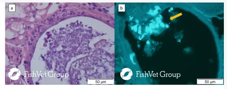
Figure 1. A section of the hepatopancreas of Pacific white shrimp Penaeus vannamei infected with Enterocytozoon hepatopenaei. (a) H & E (b) Calcofluor white streaks are E. hepatopenaei spores
(arrows). Scale: 50 µm
Diagnosis of Microsporidians primarily relies on the use of molecular diagnostic methods and light microscopy to detect spores. Visualizing Microsporidians microscopically in tissue sections can be challenging due to the very small size of the spores (~1-20µm). Special staining methods such as Trichrome, Gram, Warthin-Starry, or Calcofluor White stains can be applied. More sensitive methods like immunohistochemistry or in situ hybridization must be available for some Microsporidian species.
Various drugs have been tested for the treatment of Microsporidia infections, primarily on a research basis (e.g., benzimidazoles or fumagillin), but they show varying results across different Microsporidian species and hosts. The first successful anti-Microsporidian vaccine was developed against Loma salmonae and demonstrated the ability to reduce microsporidial fungi, indicating that this method could control Microsporidiosis.
Life cycle
Microsporidians are eukaryotic, obligate, intracellular fungal parasites that infect all animal species as well as some protists. The general life cycle of a Microsporidian can be divided into three different life stages: the infective spore stage, the proliferative stage, and the sporogonic stage. Microsporidians are primarily transmitted through aquatic environments, although vertical transmission has been demonstrated in some species. The spore is a free-living stage that can survive outside the host for many years. It has a tubular organ (Figure 2), which, when stimulated, penetrates a suitable host cell to inject sporoplasm. Once inside the host cell, the proliferative stage begins, and finally, the formation of mature spores occurs.
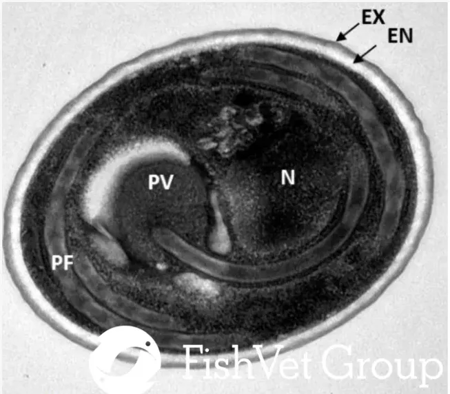
Figure 2. Transmission electron micrograph of a microsporidian spore cross-section showing nucleus (n), polar filament (PF), electron-lucent endospore wall (EN) surrounded by electron-dense exospore layer (EX), posterior vacuole (PV), and polaroplast (PP). Scale: 200 nm
Source: https://thefishsite.com/articles/microsporidians-a-macro-problem-in-aquaculture
Translated by: Trần Thị Thúy Quyên