Shrimp diseases are one of the critical issues that shrimp farmers often face. Understanding common shrimp diseases and proactive prevention measures is crucial to ensure health and increase productivity in the shrimp farming industry. In this article, we will explore some common shrimp diseases and provide suggestions on how to prevent them.
- Acute Hepatopancreatic Necrosis Disease (EMS/AHPND)
- Cause: EMS (Early Mortality Syndrome), also known as "early mortality disease," is one of the diseases causing significant losses in the shrimp farming industry. This disease is caused by a highly virulent strain of the bacterium Vibrio parahaemolyticus.
- Diagnosis: Diseased shrimp have an atrophied hepatopancreas, which appears pale to white. The intestine is empty or discontinuous, and the shrimp often have soft shells, with a high mortality rate. Black tiger shrimp with "EMS" often have a dark color, slow growth (similar to MBV stunting disease), and hepatopancreas symptoms similar to those in whiteleg shrimp, such as pale color, atrophied hepatopancreas, and empty intestines.
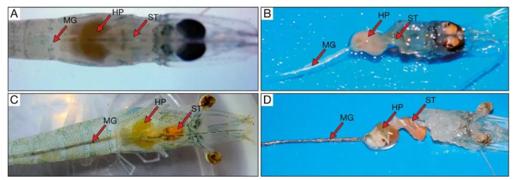
Figure 1: Whiteleg shrimp showing signs of Hepatopancreatic Necrosis Disease (A, B). The hepatopancreas (HP) is atrophied and pale; the stomach (ST) and midgut (MG) are empty. Figures (C, D) show healthy shrimp with normal-sized HP of a slightly dark orange color, and full stomach and midgut. Figures (B) and (D) are samples taken from two shrimp in figures (A) and (C) respectively. Source: Loc Tran et al., 2013.
- Prevention: To prevent EMS, apply the following measures:
- Use disease-resistant broodstock and adhere to quality monitoring procedures for postlarvae.
- Check the density of Vibrio bacteria in pond water, soil, and on postlarvae to ensure that Vibrio counts are always at a safe level.
- Polyculture with tilapia or other fish species, creating beneficial microbial communities (algae and microbes) in the pond to inhibit the growth of Vibrio bacteria (they will compete for nutrients, living environment, etc., with pathogenic bacteria). Crop rotation can be applied, with shrimp as the main crop followed by other species such as goby. To achieve sustainable and long-term shrimp farming on our land, antibiotic use should be minimized.
- White Spot Syndrome Virus (WSSV)
- Cause: White Spot Disease is one of the common diseases causing severe damage to the shrimp farming industry. There are three cases of white spot disease in shrimp with very similar external signs. This disease is caused by various factors such as bacteria, fungi, viruses, and environmental agents. For viruses, the disease is caused by white spot syndrome virus (WSSV). For bacteria, the cause is bacteria leading to Bacterial White Spot Syndrome (BWSS). Environmentally induced white spot disease is caused by high water hardness (Ca2+ and Mg2+), where shrimp absorb too much Ca2+ and Mg2+, leading to white spots on the shell.
- Diagnosis: The first step when white spot disease is detected in shrimp is to quickly identify the cause for timely treatment. PCR testing for WSSV provides fast and accurate results and should be performed immediately when shrimp show signs of white spots. If the PCR result is positive for WSSV, harvest immediately; otherwise, farming can continue, and treatment measures can be applied depending on the case.
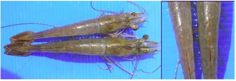
- For shrimp with White Spot Disease caused by virus: Diseased shrimp have numerous white spots measuring 0.5 – 2.0 mm appearing inside the shell, especially on the cephalothorax and the 5th and 6th abdominal segments, then spreading throughout the body. Diseased shrimp are lethargic, suddenly eat a lot then stop eating, swim sluggishly at the water surface, or gather at the pond edge. Sometimes, shrimp also show signs of red body. When white spots appear, most shrimp in the pond die (100%) within 3 - 10 days. PCR test results are positive for WSSV.
Figure 3: Whiteleg shrimp infected with WSSV.
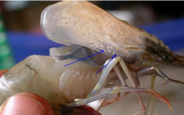
Figure 4: Black tiger shrimp infected with WSSV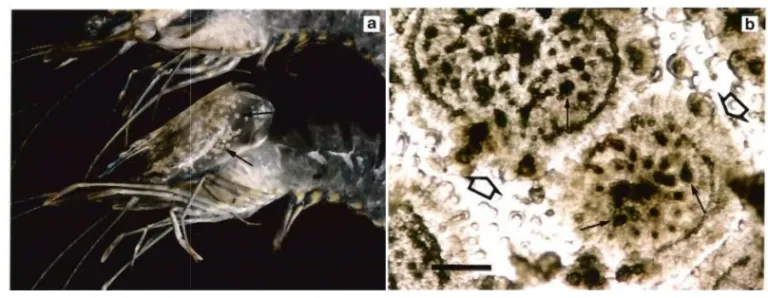
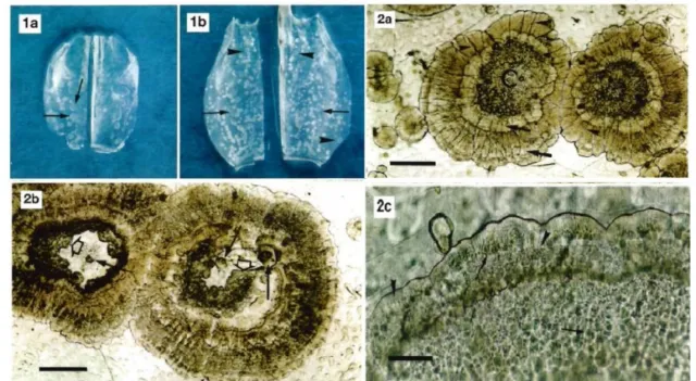
Figure 5: Microscopic examination of a fresh white spot sample showing a circular border (large, hollow arrow), with many black spots in the center.
- For shrimp with White Spot Disease caused by bacteria: Newly infected shrimp are still active, feeding and molting, and sometimes the white spots disappear after molting. With heavier infection, molting is delayed, growth is slow, and scattered deaths occur, but there is no mass mortality. Most shrimp are fouled with algae and have black gills. Diseased shrimp have opaque white spots visible on the shell throughout the body. The white spots are round, smaller, and fewer than those caused by viruses (WSSV). Microscopic examination of a fresh white spot sample shows a diffuse, lichen-like pattern with a notched circular border that is hollow in the center, whereas viral white spots have many black spots (melanin) in the center. The white spots are usually only on the outer epidermis and connective tissue. Overall, shrimp eat slower but do not suffer significant damage (Trần Việt Tiên, 2014). PCR test results are negative for WSSV.
- For shrimp with White Spot Disease caused by environmental factors: If shrimp have white spots on the cephalothorax or dorsal shell but are still healthy, with no shrimp gathering at the pond edge, and the shrimp population is still active and feeding normally, with a longer molting cycle than usual and slightly slower growth, then the white spots are caused by environmental factors, not viruses or bacteria. PCR test results are negative for WSSV.
- Prevention: To prevent white spot disease, shrimp farmers need to implement the following principles:
- Test and select WSSV-free, high-quality broodstock and postlarvae.
- Choose an appropriate farming season, avoiding stocking during cold periods.
- Control stocking density and provide sufficient oxygen to the pond.
- Water introduced into the pond must not be taken directly from nature; it must be settled and filtered.
- Prevent the entry of disease vectors such as wild crustaceans (crabs), and birds by building fences around the pond and netting to deter birds.
- Manage and closely monitor pond water quality (Trần Thị Mỹ Duyên, 2013).
- Infectious Hypodermal and Hematopoietic Necrosis Virus (IHHNV)
- Cause: Infectious hypodermal and hematopoietic necrosis virus (IHHNV) causes high mortality rates. IHHNV is classified under the family Parvoviridae, belonging to the new genus Brevidensovirus.
- Diagnosis: Whiteleg shrimp infected with IHHNV show bent or deformed rostrums, abnormal and deformed appendages in the cephalothorax region, rough and uneven shells, curled antennae, and a 10 – 30% reduction in growth, leading to stunting. For black tiger shrimp, when the disease manifests, shrimp often turn blue, the abdominal muscles become opaque white, and high mortality usually occurs 10 – 20 days after stocking. IHHNV reduces production and causes economic losses because infected shrimp at harvest are often small, uneven in size, and deformed.
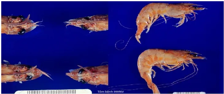
Figure 8: Whiteleg shrimp with IHHNV showing typical signs such as bent body, deformed tail, and malformation.
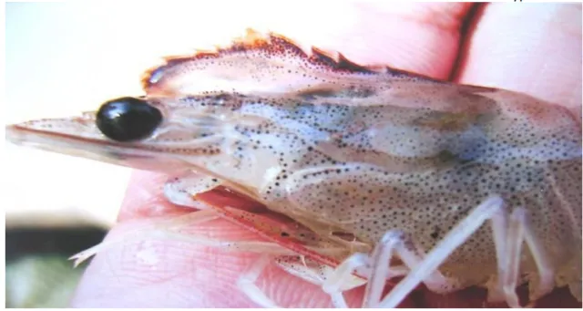
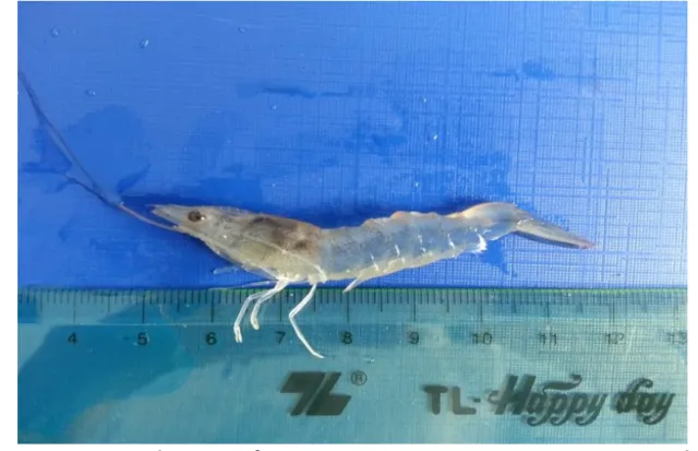
Figure 10: Whiteleg shrimp showing signs of IHHNV, with a deformed and malformed body.
- Prevention: To prevent IHHNV, shrimp farmers can implement the following measures to reduce risk:
- Use high-quality, disease-free broodstock.
- Ensure pond hygiene and remove filamentous algae, green algae, and red algae, as they can increase the risk of infection.
- Sterilization of eggs and larvae is an effective prevention method at hatchery facilities.
- Test the quality of postlarvae using PCR techniques before stocking into ponds to ensure they are free of IHHNV.
- Limit contact between ponds and maintain basic hygiene during transportation and shrimp transfer.
- Yellow Head Disease (YHV)
- Cause: The disease is caused by YHV (yellow head virus) and the gill-associated virus (GAV) complex, leading to very high mortality rates. Currently, 6 different genotypes of YHV have been identified.
- Diagnosis: Infected shrimp show yellow or brown gills, a yellow cephalothorax, a pale body, and a swollen hepatopancreas, causing the head to appear yellow. The disease causes high mortality, potentially up to 100% within 3 to 5 days of infection. PCR results are positive for YHV/GAV.
Figure 11: Black tiger shrimp dead from Yellow Head Disease YHV/GAV.
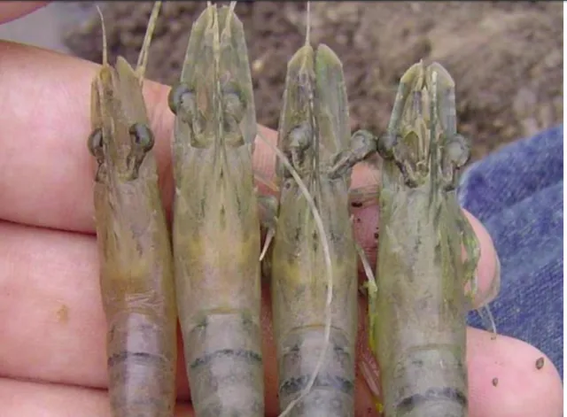
Figure 12: Close-up of the head of black tiger shrimp infected with Yellow Head Disease YHV/GAV.
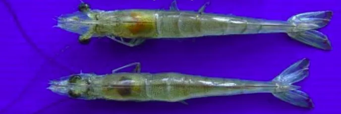
Figure 13: Whiteleg shrimp infected with Yellow Head Disease YHV/GAV (top) compared to healthy shrimp (bottom).
- Prevention: To prevent YHV, apply the following measures:
- Test the quality of postlarvae using PCR techniques before stocking into ponds.
- Maintain a clean pond environment by enhancing water quality management.
- Control stocking density appropriate for the pond volume and provide sufficient oxygen to create an unfavorable living environment for YHV.
- Limit contact between ponds and maintain basic hygiene during transportation and shrimp transfer.
- White Feces Disease (WFD/WFS)
- Cause: The exact cause has not yet been determined. Many studies suggest that white feces disease in shrimp is caused by Vibrio bacteria, while other studies attribute it to gregarines or a group of parasites called Vermiform. Some research indicates that infected shrimp show the presence of various pathogens such as bacteria (Vibrio group), parasites (Vermiform, gregarines), and viruses.
- Diagnosis: Diseased shrimp excrete white feces; occasionally, the fecal strands are also pale yellow. The hepatopancreas is atrophied or soft, and shrimp with white feces disease often exhibit soft shells or loose shells. A few days after infection, shrimp become weak and swim sluggishly at the water surface, gradually weakening and dying. Simple tests should be conducted to accurately identify the cause of the disease for appropriate treatment. The first and simplest step is to check for parasites in the shrimp intestine by cutting a section of the intestine and examining it under an optical microscope. If no parasites are found, proceed to check the total Vibrio bacteria in the pond environment. If the Vibrio count is too high, the cause may be Vibrio bacteria.
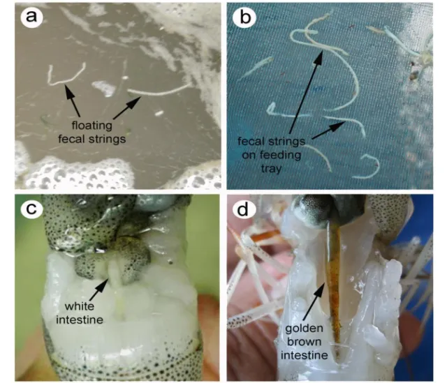
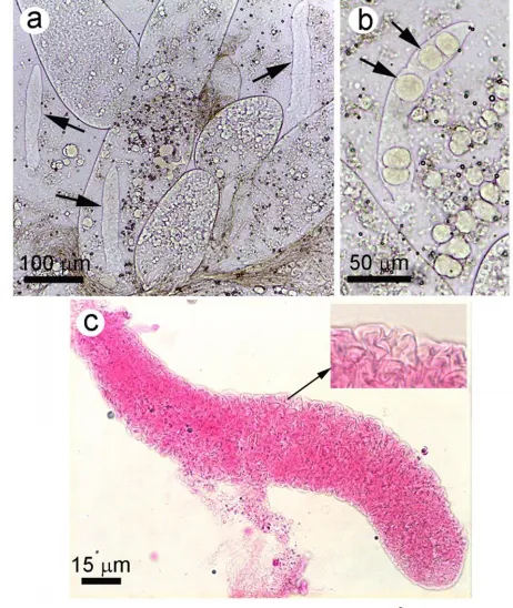

Figure 16: H&E stained shrimp hepatopancreas tissue sample clearly showing the morphology of vermiform and spore-like structures.
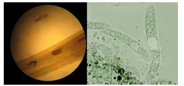
Figure 17: Gregarines isolated from the intestine of shrimp with white feces disease.
- Prevention: To prevent this disease, shrimp farmers need to apply the following integrated prevention measures:
- Carefully manage water quality and control shrimp feed.
- Control stocking density and provide sufficient oxygen to the pond. This reduces the organic matter content at the pond bottom and inhibits the growth of Vibrio spp. bacteria.
- Use microbial products to control bacteria and limit the growth of pathogenic Vibrio spp.
- Polyculture of shrimp with tilapia also effectively controls the growth of Vibrio bacteria in the pond.
- To control gregarines, use garlic at a dosage of 5 - 10g/kg of feed for high effectiveness and periodically deworm shrimp.
- Taura Syndrome
- Cause: The disease is caused by taura syndrome virus (TSV).
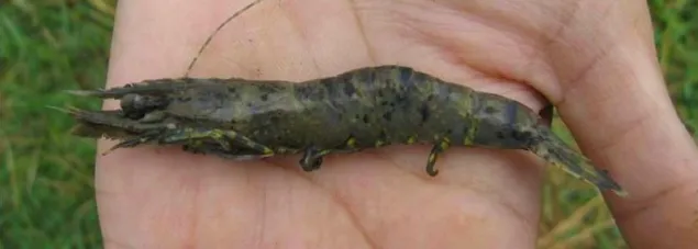
- Diagnosis: Whiteleg shrimp infected with TSV have a pale red color, especially in the tail region. Additionally, shrimp show other signs such as soft shells and empty intestines. Taura syndrome causes high mortality (typically 40% to 90%) and spreads rapidly. Taura virus can infect black tiger shrimp, causing red tail disease: shrimp have a red color throughout the tail fan and subsequent body segments extending towards the head; walking legs and swimming legs also appear red.
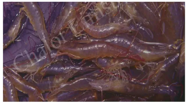
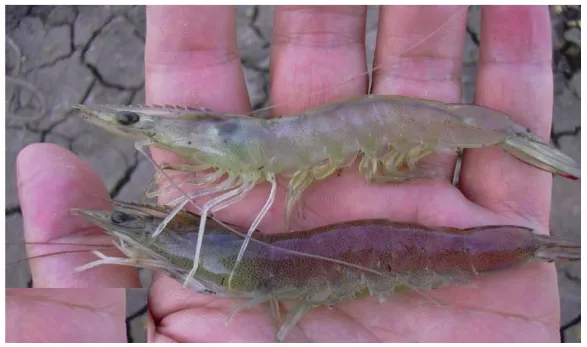
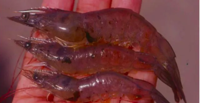
Figure 20: Whiteleg shrimp with Taura Syndrome, accompanied by symptoms of bacterial infection.
- Prevention: To prevent this disease, shrimp farmers can implement the following integrated prevention measures:
- Test and select SPF (Specific Pathogen Free), high-quality broodstock and postlarvae.
- Check water quality and maintain a stable water environment, including appropriate temperature, pH, and oxygen levels.
- Choose an appropriate farming season, avoiding stocking during cold periods.
- Control stocking density and provide sufficient oxygen to the pond.
- Water introduced into the pond must not be taken directly from nature; it must be settled and filtered.
- Prevent the entry of disease vectors such as wild crustaceans (crabs), and birds by building fences around the pond and netting to deter birds.
- Muscle Necrosis, White Tail, Opaque Muscle Disease
There are many causes of muscle necrosis, white tail, and opaque muscle disease in shrimp. The general sign of this disease is that the tail muscle or muscles in other body segments, or the entire body, turn white or opaque and show signs of necrosis. The causes/cases of opaque muscle in shrimp are as follows:
- Opaque muscle combined with bent body: This usually occurs when lifting feeding trays or nets out of the water during the day when temperatures are very hot. Shrimp jump and flick strongly, and then some become bent-bodied.