While classical R&D continues, is the use of AI a good direction for the future?
Occurring singly or as co-infections, current disease threats including Enterocytozoon hepatopenaei (EHP), Acute Hepatopancreatic Necrosis Disease (AHPND), White Feces Syndrome (WFS), and White Spot Syndrome Virus (WSSV) remain a productivity threat to many farms in Asia. Added to this is Infectious Myonecrosis Virus (IMNV), prevalent in Indonesia. What can be done to reduce co-morbidity and track disease hotspots, maintain consistency, and improve predictability in aquaculture? This is key to living with disease and maintaining productivity.
Following two presentations updating research on EHP, the use of AI, and Data Analytics to predict disease hotspots, Dr. Daranee Seguin, Thailand, chaired a workshop at TARS 2023 to provide some answers. Industry participants in the workshop were Dr. Lộc Trần, Founder and Director, ShrimpVet Laboratory Vietnam, and Dan Fegan, Group Director, SyAqua, Thailand.
EHP Management
There continues to be a frequent occurrence of Enterocytozoon hepatopenaei (EHP) outbreaks causing hepatopancreatic microsporidiosis (HPM).
Dr. Kallaya Sritunyalucksana provided the latest update from laboratory studies in Thailand with EHP. Kallaya is the Principal Researcher of the Aquatic Animal Health Research Team at the National Center for Genetic Engineering and Biotechnology (BIOTEC), National Science and Technology Development Agency (NSTDA).
The research team at BIOTEC has studied the life cycle, transmission routes, and virulence mechanisms of this microsporidian since 2009 to provide recommendations for shrimp farmers in controlling it.
A cohabitation model was used to answer some farmer questions. EHP is transmitted horizontally, not vertically.
“When we added infected shrimp to unstocked shrimp tanks in the lab, infection occurred within 14 days. Spores are present in water and feces. Infection is transmitted orally, and the initial infection is gastrointestinal. Naturally, the infection rate is very high. Stocking density increases the spread of EHP,” said Kallaya.
Results from the cohabitation model and EHP spore cleaning studies have helped her team design EHP control strategies such as using the spore wall protein (SWP)-PCR detection method (aroenlak et al., 2016), pond preparation by high pH treatment, low temperature for live feed preparation, and detection of mechanical EHP carriers such as bivalves, etc.
“Together with CEFAS and the University of Exeter in the UK, we sequenced the entire genome using purified EHP spores and submitted it to GenBank (Boakye et al., 2016).
We know that the EHP genome contains 4 ATP transporter genes but has lost 8 of the 10 essential glycolytic genes for ATP production. With the downregulation of these ATP transporter genes, EHP cannot replicate. This implies that this parasite is entirely dependent on receiving ATP from the host, leading to slow shrimp growth.”
Steps to Mitigate EHP
Disinfectants such as KMn04 15ppm, 60% active chlorine, and 20% ethanol inhibit EHP spores. The post-harvest recommendation is to treat contaminated water and let it sit for at least 10 days to inactivate EHP spores.
Freezing at -20°C can inactivate spores. This is crucial when using live feeds such as artemia and polychaetes. Related to feed production is the infectivity of spores being inhibited at 75°C within 1 minute (Figures 1 and 2). Spores treated at 75°C and untreated (28°C) were experimentally added to pelleted feed and fed to shrimp. It was found that shrimp fed with spores treated at 75°C showed no EHP multiplication using qPCR, while shrimp fed with untreated spores showed high EHP multiplication. Considering the level of heat exposure during feed processing (at or exceeding 75°C for 60 seconds), such feeds should be considered non-risky for EHP transmission.
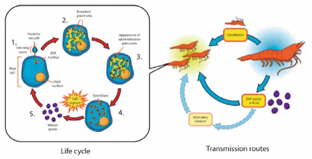
Figure 1. Diagram illustrating the life cycle and transmission route of EHP. Photo by Chaijarasphong et al., 2020
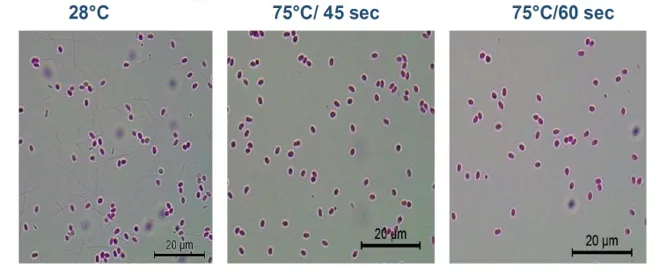
Figure 2. EHP spore density in cells at 75°C at 45 seconds and 60 seconds compared to 28°C
HPM Diagnosis
Attention is drawn to this issue when farmers look for spores in tissues and feces. “HPM has no clear symptoms for convenient diagnosis. It is suspected when there is slow growth. Histological diagnosis depends on the presence of spores in the hepatopancreas. The spores are very small, and with a microscope, a 100X objective lens is needed. Sometimes spores appear in small numbers, even with severe infection,” said Kallaya. She added that a high density of spores in the hepatopancreas indicates a severe EHP infection.
Stemming from EHP genome research, BIOTEC has developed a specific PCR method to detect the EHP spore wall protein (SWP) gene (SWP-PCR). It is recommended for detecting EHP in shrimp samples and in feces, feed, and environmental samples for potential EHP carriers, along with histology, to avoid false positives. The important message is that post-larvae (PL) shrimp should be tested using SWP-PCR at least twice within a 7-day interval (PL5 and PL12) before stocking. A portion of the PL5 tested first must be cultured under stress conditions for 7 days to allow EHP to multiply. Screening should be performed again at PL12 or before stocking to ensure the shrimp are EHP-free.
EHP Carriers
Mechanical EHP carriers have been identified as the mussel Mytilopsis leucopheata.
EHP cannot replicate in this mussel species but can survive in its digestive tract. A cohabitation model of shrimp and mussels with spores in their digestive glands showed that shrimp became infected after 10-20 days. Other pathogen carriers include artemia, polychaetes, and other bivalve species. It is recommended to remove bivalves and thoroughly wash artemia and polychaetes before use.
EHP is a Partial Cause of WFS
“We now know that EHP is not the cause of White Feces Syndrome (WFS) but is a partial cause when combined with bacteria. EHP-WFS is associated with 6HP (low toxin-producing Vp-AHPND). Widespread bacterial infection in EHP-infected shrimp can lead to WFS and mortality. EHP infection leads to a higher susceptibility to 6HP,” Kallaya indicated as shown in Figure 3.
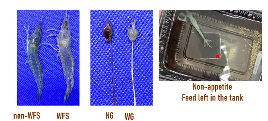
Figure 3. WFS reproduction in the EHP-WFS laboratory model. Data source: Update on microsporidian, Enterocytozoon hepatopenaei (EHP), presented by Kallaya Sritunyalucksana, TARS 2023

An important message from Kallaya Sritunyalucksana is: “Post-larvae shrimp should be tested using SWP-PCR at least twice within a 7-day interval (PL5 and PL12) before stocking.”
WFS results from the sloughing, degradation, and release of HP cells from the tubule epithelium. Recovery can be faster if detected early. BIOTEC has an EHP-WFS laboratory model that can allow testing of inhibitors or WFS management practices.
Source: Aquaculture Asia Pacific
Translated by: Trần Thị Thúy Quyên